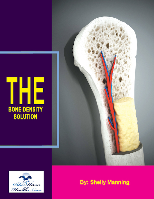
Bone Density Solution By Shelly Manning As stated earlier, it is an eBook that discusses natural ways to help your osteoporosis. Once you develop this problem, you might find it difficult to lead a normal life due to the inflammation and pain in your body. The disease makes life difficult for many. You can consider going through this eBook to remove the deadly osteoporosis from the body. As it will address the root cause, the impact will be lasting, and after some time, you might not experience any symptom at all. You might not expect this benefit if you go with medications. Medications might give you some relief. But these are not free from side effects. Also, you will have to spend regularly on medications to get relief from pain and inflammation.
How do healthcare providers determine the severity of osteoporosis?
Healthcare providers make the determination of the degree of osteoporosis primarily based on bone density, clinical, and evaluation of one’s risk factors. This is how they gauge the condition:
1. Bone Density Testing (DEXA Scan)
The most frequent test of bone mineral density (BMD) is a Dual-Energy X-ray Absorptiometry (DEXA or DXA) scan. It produces a T-score, which is helpful in estimating the severity of osteoporosis:
Normal bone density: T-score ≥ -1.0
Osteopenia (low bone density): T-score -1.0 to -2.5
Osteoporosis: T-score ≤ -2.5
Severe osteoporosis: T-score ≤ -2.5 accompanied by a history of one or more fragility fractures (fractures resulting from minimal trauma, e.g., fall from standing height).
2. Fracture History
One of the most significant elements in determining osteoporosis severity is the history of fractures, particularly fragility fractures, that are common in people with weakened bones.
Spinal, hip, or wrist fractures are especially ominous indications and are evidence of more advanced osteoporosis.
Fractures are evidence of extreme loss of bone, and osteoporosis is staged on the basis of the frequency and location of fractures.
3. Clinical Risk Assessment
Clinicians use a risk assessment tool such as the FRAX (Fracture Risk Assessment Tool) to estimate the 10-year probability of fracture based on factors such as:
Age, sex, weight, height
History of prior fractures
Family history of osteoporosis or fractures
Smoking and alcohol use
Use of drugs with bone-destabilizing effects (e.g., corticosteroids)
Such diseases as rheumatoid arthritis or kidney disease
This guides the severity of the disease and treatment options.
4. Spinal Imaging
In some cases, medical providers may use X-rays or MRI to assess vertebral fractures (common in osteoporosis), particularly if a patient comes in with back pain or other symptoms suggestive of a spinal condition.
Vertebral fractures can be asymptomatic but contribute to overall bone frailty and increased risk of fractures.
5. Physical Examination and Symptoms
While physical examinations alone do not diagnose the severity of osteoporosis, they can help identify signs of bone fragility, such as loss of height or kyphosis (hunched shoulder), both of which are frequently associated with vertebral fractures and severe osteoporosis.
Symptoms like back pain (especially after lifting, bending, or a fall) may indicate vertebral fractures or widespread bone loss.
6. Bone Turnover Markers (Blood or Urine Tests)
Although not employed as an integral component of standard care for the diagnosis or determination of severity of osteoporosis, markers of bone remodeling can be utilized to assess bone remodeling rate.
Elevation of bone resorption markers (e.g., C-terminal telopeptide, or CTX) and reductions in markers of bone formation (e.g., osteocalcin) can signal active bone loss, in agreement with more extensive osteoporosis.
7. General Health and Comorbidities
Comorbid illness (e.g., diabetes, rheumatoid arthritis, hyperthyroidism) can influence osteoporosis severity and progression.
In the event of a person having other conditions influencing bone status or healing, severity of osteoporosis may be rated higher.
8. Risk of Future Fractures
The risk of fracture in the future is one consideration for how severe osteoporosis is. Healthcare professionals may apply the FRAX score or another clinical assessment to predict the 10-year fracture risk and make a prediction based on it about the severity of osteoporosis.
The higher the future risk of fracture, the earlier prevention and treatment must be administered.
Treatment by Severity
Mild to moderate osteoporosis: May be managed with lifestyle change (e.g., exercise, vitamin D and calcium supplementation) and medication like bisphosphonates (e.g., alendronate) or selective estrogen receptor modulators (SERMs).
Severe osteoporosis: May be managed with intensified treatment, e.g., stronger drugs (e.g., denosumab, teriparatide) and more frequent check-ups, and fracture prevention (e.g., measures to prevent falls).
Would you like more information on treatment or osteoporosis management at different stages?
A physical examination is a critical component of osteoporosis diagnosis but cannot diagnose osteoporosis independently. Osteoporosis is a condition in which the bone mass is reduced and the bones are made weaker, putting the individual at risk of fractures. A physical examination is required because:
1. Identification of Symptoms and Risk Factors
Clinical Observations: Osteoporosis is not always symptomatic, but physical examination can detect signs that may signal osteoporosis. These are:
Postural changes, such as kyphosis (spinal curvature or a “dowager’s hump”), that result from vertebral fractures.
Loss of height: Progressive loss of height over time could represent spinal fractures or compression fractures due to weakened bone.
Tenderness or pain: Areas of the body may be tender, especially in the hips or spine, which may suggest fractures or weakened bones.
2. Assessing Balance and Risk of Falling
A physical exam allows medical providers to assess balance and mobility. Patients with osteoporosis are at increased risk for falling because they have decreased bone density and muscle weakness. Assessing such factors as:
Posture: Abnormal posture or spinal deformities may be an indication of vertebral compression fractures.
Coordination and gait: Weakness or unsteadiness of gait increases the risk of falls, which lead to fractures in individuals with osteoporosis.
Evaluation of the risk of falls helps clinicians to prescribe preventive measures, such as home modifications or physical therapy.
3. Fracture or Deformity Detection
A physical exam can validate the existence of fracture signs that can be the result of weakened bones:
Spinal tenderness: Palpation of the spine will reveal tenderness in the areas where vertebral fractures may have occurred.
Hip or wrist pain: Common areas for fractures among osteoporosis patients, particularly after an accident or fall.
Bone deformities: Deformities of the arms, legs, or spine (such as a collapsed vertebra) may be suggestive of long-standing osteoporosis and may help choose locations where further imaging may be appropriate.
4. Establishing Baseline Data to Track
Physical examination provides useful baseline data for assessing trends in a patient’s status over the course of time. For example, an alteration in posture or loss of height over months or years can be utilized to follow the course of osteoporosis and guide treatment.
5. Establishing Coexisting Conditions
Osteoporosis also often exists with other diseases, such as arthritis, muscular weakness, or malnutrition, which may affect the bone. Comorbid conditions are diagnosed on physical examination to affect osteoporosis treatment, such as deficiency of vitamin necessary for bone density.
6. Osteoporosis and Other Conditions
Physical exam can also be utilized to differentiate osteoporosis from other diseases that can produce similar symptoms (e.g., degenerative disc disease, osteomalacia, or rheumatoid arthritis). Clinical findings, as well as patient history, can direct healthcare providers to appropriate testing.
While a physical examination cannot diagnose osteoporosis on its own, it provides essential information to aid in the assessment of symptoms, risk factors, and fall risk as well as fracture or deformity due to bone loss. It also serves as the foundation for further diagnostic testing, such as bone mineral density testing (DXA scan), to establish the diagnosis and guide treatment.
Would you like to learn more about what’s next with diagnosing osteoporosis following a physical examination?

Bone Density Solution By Shelly Manning As stated earlier, it is an eBook that discusses natural ways to help your osteoporosis. Once you develop this problem, you might find it difficult to lead a normal life due to the inflammation and pain in your body. The disease makes life difficult for many. You can consider going through this eBook to remove the deadly osteoporosis from the body. As it will address the root cause, the impact will be lasting, and after some time, you might not experience any symptom at all. You might not expect this benefit if you go with medications. Medications might give you some relief. But these are not free from side effects. Also, you will have to spend regularly on medications to get relief from pain and inflammation.